Diagnosing instability of ligamentous syndesmotic injuries: A biomechanical perspective. Review published in Clinical Biomechanics
18/03/2021
Spennacchio P., Seil R., Gathen M., Cucchi D. Diagnosing instability of ligamentous syndesmotic injuries: A biomechanical perspective, Clinical Biomechanics, Volume 84, 2021, 105312, ISSN 0268-0033, https://doi.org/10.1016/j.clinbiomech.2021.105312.
Highlights
- Different experimental setups available to investigate syndesmotic stability.
- Ex vivo models: significant kinematic role of anterior inferior tibiofibular ligament.
- Isolated anterior inferior tibiofibular ligament lesion can cause dynamic instability.
- Suspected syndesmotic instability: 2nd level diagnostics, minimize risk for sequelae.
- Computed tomography can improve the diagnosis of syndesmotic instability.
Abstract
Background: High ankle sprains are insidious injuries associated with a long recovery period, functional impairment and long-term sequelae if mistreated. This systematic review investigates the biomechanical knowledge on the kinematic consequences of sequential syndesmotic ligamentous injuries, aiming to furnish an updated and objective contribution for the critical appraisal and further elaboration of current diagnostic algorithms for high ankle sprains.
Methods: A systematic review was performed to identify human biomechanical studies evaluating the stabilizing role of the syndesmotic ligaments. Special attention was paid to identify the smallest lesion within the progressive simulated injuries able to provoke statistically significant changes of the syndesmotic kinematic on the specimen, the mechanical solicitation that provoked it, and the measurement methodology.
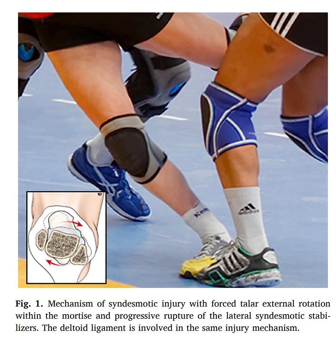
Findings: Fourteen studies were included. In eight articles already an isolated injury to the anterior inferior tibiofibular ligament provoked significant changes of the syndesmotic kinematic, which was always depicted under an external rotation torque. In three articles an isolated deltoid ligament injury provoked significant changes of the syndesmotic kinematic. Four articles described a direct measure of the bony movements, whereas seven collected data through conventional radiography or CT-scan imaging and three via a 3D motion analysis tracking system.
Interpretation: An isolated lesion of the anterior inferior tibiofibular ligament can provoke significant kinematic modifications in ex vivo syndesmotic models and may be responsible of subtle patterns of dynamic instability, regardless of further syndesmotic ligamentous injuries. The data observed support efforts to define reliable CT imaging parameters to improve non-invasive diagnostic of subtle forms of syndesmotic instability.